Numb Fingers: Which Cervical Level Is the Cause? The Answer Isn't What You'd Expect
By Yutoku Yakuhin··7 min read
Adapted from a Chinese-language rehabilitation explainer. For general education only — not a diagnosis. Numb fingers have many possible causes; please see a qualified clinician for an in-person examination.
The Short Answer, With a Cold Shower First
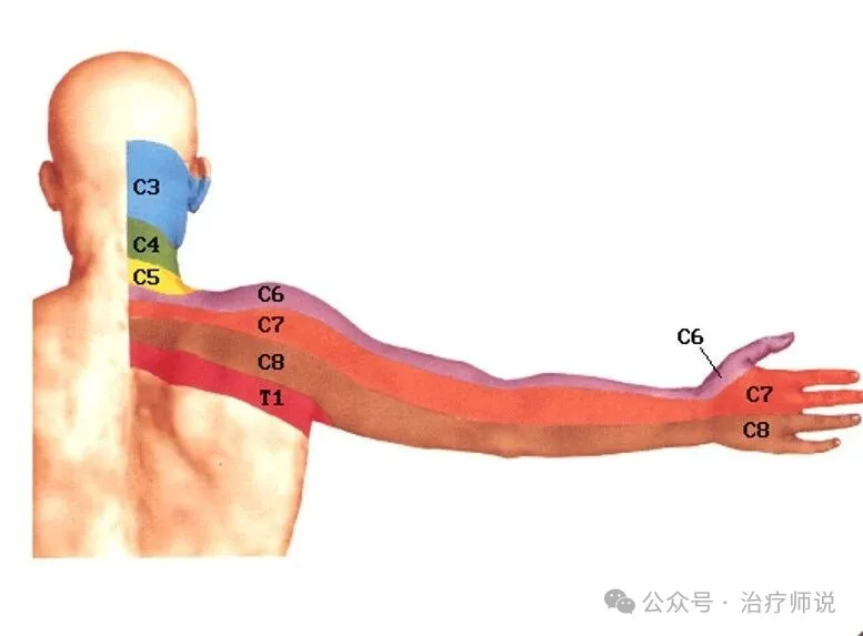
A chart goes around online: thumb and index finger numb = sixth cervical vertebra; middle finger = seventh; ring and little finger = eighth.
The broad direction is right. But reading it like a lookup table is a reliable way to end up with the wrong conclusion, for three reasons.
1. "Which cervical vertebra?" is not quite the right question. The neck has 7 vertebrae (C1–C7), but 8 pairs of nerve roots leave it (C1–C8). The C8 root exits between C7 and T1 — below the last cervical vertebra, yet there is no "eighth cervical vertebra." That's why clinicians talk about the C6 nerve root or the C7 nerve root, not "the sixth bone."
2. One level affects a region, not a single finger. A disc herniation at C5–C6 usually compresses the C6 root, because each cervical root exits above the vertebra of the same number (the C6 root leaves between C5 and C6). So "a problem at the sixth vertebra" really has to be translated into "C5–C6 level → C6 root" to be accurate.
3. Most importantly, numb fingers aren't always a neck problem. Carpal tunnel syndrome, ulnar nerve entrapment, diabetic peripheral neuropathy, and thoracic outlet syndrome can all numb the fingers. Diagnosing the neck from "which finger is numb" alone is one of the most common clinical mistakes.
So instead of a quick-lookup chart, here is the logic clinicians actually use.
1. Start With the Map: Nerve Root to Fingers
This is the foundation of clinical localisation, based on the standard dermatome maps and consistent with the American Academy of Family Physicians neurological localisation table.
| Nerve root | Main sensory area | Fingers usually affected | Typical level |
|---|---|---|---|
| C5 | Outer upper arm, shoulder | Not usually the fingers on its own | C4–C5 |
| C6 | Outer forearm, thumb | Thumb and index finger | C5–C6 |
| C7 | Back of the forearm, middle finger | Middle finger (may spread to index or ring finger) | C6–C7 (most common) |
| C8 | Inner forearm | Ring and little finger | C7–T1 |
| T1 | Inner forearm, small muscles of the hand | Inner edge of the little and ring finger | T1–T2 |
One line to remember: C6 = thumb and index, C7 = middle, C8 = ring and little.
C7 is the nerve root most often compressed, followed by C6. That's why "middle finger numbness" is the pattern clinics see most.
2. Why You Can't Just Use the Chart: Three Rules
A dermatome map can narrow things down. It can't confirm anything. Real localisation needs several pieces of evidence to agree.
Rule 1: Sensation, strength, and reflex must point to the same nerve
Sensation is only one-third of the picture. A clinician checks all three:
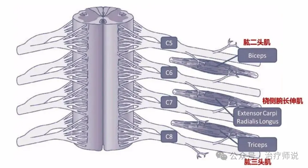
| Nerve root | Sensation | Key muscle strength (movement) | Reflex |
|---|---|---|---|
| C5 | Outer upper arm | Lifting the arm sideways (deltoid) | Biceps |
| C6 | Thumb, index finger, outer forearm | Extending the wrist | Brachioradialis |
| C7 | Middle finger, back of forearm | Straightening the elbow (triceps), bending the wrist | Triceps |
| C8 | Ring and little finger, inner forearm | Bending the fingers | Usually no distinct reflex |
The key point: if only the thumb is numb, but wrist extension is strong and the brachioradialis reflex is normal, the C6 explanation is in doubt. When the three don't line up, the numbness may be coming from somewhere else.
Rule 2: The pattern should be a band, not a spot
Numbness from a compressed nerve root usually follows a continuous strip: neck or shoulder → upper arm → forearm → fingers.
Carpal tunnel numbness is different. It sits in the thumb, index, middle, and thumb-side half of the ring finger, tends to be worse at night, and can be reproduced by tapping over the wrist (Tinel's sign). It does not travel up the arm.
Rule 3: An MRI can't convict on its own
A disc bulge on MRI doesn't prove it's causing your symptoms. Plenty of people with no symptoms at all have bulges on imaging. Clinically, the symptoms, the examination findings, and the imaging all have to agree before a level is considered responsible.
3. So How Do You Match It Up? Four Common Patterns
These are ways to narrow it down, not conclusions.
Pattern 1 — Thumb and index finger numb
- Most likely: C6 root (C5–C6 level).
- Often with: numbness down the outer forearm, weak wrist extension, a weaker brachioradialis reflex.
- Rule out first: carpal tunnel syndrome — waking at night with numb hands, relief from shaking them out, weak thumb opposition. It's the condition most easily confused with a neck cause.
Pattern 2 — Middle finger numb (sometimes with the index finger)
- Most likely: C7 root (C6–C7 level), the most common of all.
- Often with: weak elbow extension, a weaker triceps reflex, numbness on the back of the forearm.
Pattern 3 — Ring and little finger numb
- Most likely: C8 root (C7–T1 level) or T1.
- Often with: weak finger bending, wasting of the small hand muscles (the fleshy pads at the thumb and little-finger sides flatten).
- Rule out first: ulnar nerve entrapment — numbness when tapping the groove at the elbow, a claw-like hand. This is another frequent look-alike.
Pattern 4 — Both hands numb, glove-like and symmetrical
- Be alert for: diabetic peripheral neuropathy, cervical canal stenosis, or a whole-body cause.
- This doesn't match any single nerve root, and checking only the neck would miss it.
4. More Important Than "Which Level": Red Flags
Localising the level is the clinician's job. Red flags are something everyone should know. If any of these appear, don't watch and wait, and don't try massage first — go to a hospital.
- Clumsy hands, trouble with buttons or chopsticks → possible cervical myelopathy (spinal cord compression).
- Unsteady walking, a "walking on cotton" feeling, tripping easily → possible spinal cord compression. Needs prompt attention.
- Bladder or bowel changes (urgency, incomplete emptying, incontinence) → possible cord compression emergency.
- Numbness or weakness in the legs too → likely more than a single nerve root problem.
- Numbness that keeps getting worse, or muscles wasting → the nerve damage is progressing.
- Numbness after neck trauma → fracture or dislocation has to be excluded.
Important: with myelopathy, forceful neck manipulation or "cracking" can cause serious harm. Don't head to a massage or bone-setting shop for numb hands. Get a diagnosis first.
5. Three Practical Tips for Heavy Phone and Screen Users
- Find out first, then treat. There are at least five or six causes of hand numbness. Start with a physical examination and, if needed, nerve conduction studies plus a cervical MRI. Don't guess.
- Don't stay in one posture. Every 30–45 minutes, get up and move your neck and shoulders, and raise the screen to eye level. Looking down can multiply the load on the cervical spine several times over.
- Train the deep neck muscles; don't whip the head around. Fast, wide neck circles aren't always kind to a spine that already has degenerative changes. Gentle isometric stabilisation work is safer. If you already have symptoms, talk to a professional first.
Ask the Right Question
"My fingers are numb — which vertebra is it?"
The honest answer is that "which finger" can't settle it on its own. It takes sensation, strength, reflexes, provocation tests, and imaging fitted together to point at a nerve root, and then the nerve root has to be translated into a disc level.
So next time your hands go numb, don't scare yourself with a chart online, and don't rush to be massaged. Find a clinician who will do a full neurological examination. It's the fastest and cheapest route to an answer.
References: Eubanks JD. Cervical Radiculopathy: Nonoperative Management of Neck Pain and Radicular Symptoms. Am Fam Physician. 2016;93(9):746–754 (nerve-root localisation table); and consensus guidance on cervical spondylosis that requires symptoms, examination findings, and imaging to agree before identifying the responsible level. This article is educational and is not a diagnosis.